Imaging Gallery
Explore Our Center
-
 Holiday Party 2022.
 -
 AHA 2022.
 -
 AHA 2022.
 -
 Cathedral of Learning.
 -
 Discussion in the conference room.
 -
 Acoustic field calibration.
 -
 Multi-modality biomedical ultrasound imaging.
 -
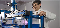
 Microbubble fabrication.
 -
 Rabbit femoral artery sections before (upper panels) and 6 weeks after (middle and lower panels) balloon injury.
 -
 Contrast ultrasound of the rabbit femoral arteries before (A, B) and 6 weeks after (C, D) balloon injury.
 -
 Ultrasound radiation force-mediated delivery of stem cells to vascular targets.
 -
 The MSCs adherent to the aortic endoluminal surface after ultrasound mediated delivery survive and undergo morphological changes.
 -
 Postmortem histology of tumors in mice after intravenous delivery of pCMV-TK (a, c, d) or pEGFP-C1 (b)-loaded microbubbles and treatment with ultrasound and ganciclovir. (a, b).
 -
 Growth of murine tumors after intravenous injection of either pCMV-TK (□) or pEGFP-C1 (▲)-loaded microbubbles and treated with ultrasound.
 -
 In vitro model of microvascular embolization and sonoreperfusion.
 -
 Quantification of sonothrombolysis effects. Blood clot volume derivation from an optical coherence tomography (OCT) scan and 3-D rendering of thrombus.
 -
 Quantification of microbubble attachment of mesenchymal stem cells (MSCs) using flow cytometry.
 -
 Acoustic radiation force on microbubbles can be used to delivery stem cells for vascular cell therapy.
 -
 3D tumor volumes in mice receiving UTMD-mediated delivery of control siRNA (top) or UTMD-mediated delivery of EGFR–siRNA (bottom).
 -
 The UPMC Cam, the ultra-fast imaging system for studyng the dyanmic behavior of microbubbles.
 -
 Overview of the UPMC Cam. This system is capable of imaging microscopic bright-field and fluorescence movies at 25 million frames per second for 128 frames.
 -
 Optical design of the UPMC Cam.
 -
 Sequential frames of a bright-field movie of lipid microbubbles under ultrasound excitation (f= 2.25 MHz, Pa = 1.0 MPa), demonstrating ultrasound induced microbubble vibration and breaking.
 -
 Sequential bright-field high-speed microscopic images of a microbubble adjacent to a thrombus during ultrasound delivery (1 MHz, 1.5 MPa).
 -
 Sequential bright-field high-speed microscopic images of multiple microbubbles (arrowheads in frame 7) adjacent to a thrombus (arrow in frame 7) during ultrasound exposure.
 -
 The dual-mode US system for US-TS Imaging.
 -
 Contrast enhanced ultrasound imaging using microbubbles targeted to E-selectin can be used for ischemic memory imaging.
 -
 Immunofluorescent staining of post-ischemic (A-C) and nonischemic (D-F) myocardium 4 hours after reperfusion.
 -
 Nonlinear intravascular ultrasound imaging with microbubble based contrast agents.
 -
 In vivo model of sonoreperfusion. Contrast ultrasound images from control (top) and treatment (bottom) animals at each of four experimental stages.
 -
 Hematoxylin and eosin-stained treated (c,d) and control(a,b) rat hindlimb muscle. The rats that received treatment had less microvascular obstruction than controls.
 -
 High-speed fluorescence recordings with the UPMC Cam reveal a variety of microbubble coating behaviors not evident from bright-field recordings.
 -
 High-speed fluorescence recordings with the UPMC Cam reveal a variety of microbubble coating behaviors not evident from bright-field recordings.
 -

-

-

-

-

-

-

-

-

-

-

-

-

-

-

-

-

-

-