High Speed Imaging
We are developing a high-speed microscopy imaging system capable of acquiring images at up to 25 million frames per second under either brightfield or fluorescent light conditions (Chen X, Wang J, Versluis M, de Jong N, and Villanueva FS, Ultra-fast bright field and fluorescence imaging of the dynamics of micrometer-sized objects. Rev Sci Instrum, 2013. 84(6): p. 063701). Such a system allows us to directly visualize the mechanics of a single oscillating microbubble or the dynamics of a microbubble’s interaction with another object or environment. This tool, the only one of its kind in the world, will help us better understand microbubble dynamics, mechanisms of microbubble bioeffects, and improve their design for specific applications. Our ultra fast imaging system may also have applications to imaging phenomena in other areas such as cell biology and microbiology that may occur at time scales smaller than ever before realized.
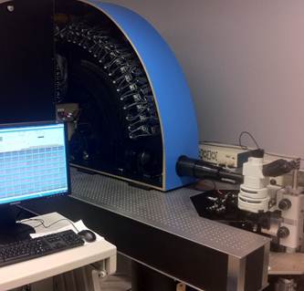
A photograph of UPMC Cam, the ultra-fast imaging system for studyng the dyanmic behavior of microbubbles.
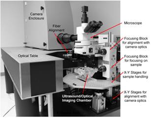
Overview of UPMC Cam. This system is capable of imaging microscopic bright field and fluorescence movies at 25 million frames per second for 128 frames, with a frame size of 920×616 pixels. Adapted from Chen X,…Villanueva FS , Rev Sci Instrum 2013; 84.
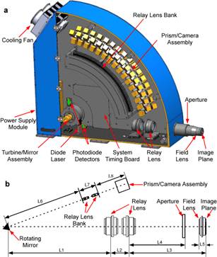
Optical design of the UPMC Cam. For clarity, all USB hubs and relay circuits were removed from this drawing. Adapted from Chen X,…Villanueva FS , Rev Sci Instrum 2013; 84.
This is a bright field movie of lipid microbubbles under ultrasound excitation (f=2.25 MHz, Pa=1.0 MPa), demonstrating ultrasound-induced microbubble vibration and breaking. Imaging is at 25 million fps, and playback is at 30 fps. Frame size is 27 µm × 27 µm. Adapted from Chen X,…Villanueva FS, Rev Sci Instrum 2013; 84.
This is a bright field movie of lipid microbubbles under ultrasound excitation (f=2.25 MHz, Pa=0.5 MPa), demonstrating ultrasound-induced microbubble oscillation. Imaging is at 25 million fps, and playback is at 30 fps. Frame size is 22 µm × 22 µm.
This is a brightfield movie of a microbubble oscillating adjacent to a thrombus (left) under the influence of ultrasound (1 MHz, 1.5 MPa), causing intermittent thrombus deformation. After the microbubble has disappeared from the microscopic field, a “pit” remains in the thrombus at the previous site of microbubble oscillation. Imaging is at 5 million fps, and playback is at 8 fps. Frame size is 55 µm x 55 µm. Adapted from Chen X, …Villanueva FS, Ultrasound Med Biol 2014;40.
This is a brightfield movie of multiple microbubbles oscillating adjacent to a thrombus (left) during ultrasound exposure (1 MHz, 1.5 MPa). Acoustic radiation force causes oscillating microbubbles to migrate towards the thrombus (a new microbubble enters the field of view from the right). Note the marked cyclic thrombus deformation in response to ultrasound-induced microbubble oscillations compressing the thrombus, and ultimate microbubble destruction or formation of smaller microbubbles. Imaging is at 5 million fps, and playback is at 8 fps. Frame size is 55 µm x 55 µm. Adapted from Chen X,…Villanueva FS , Ultrasound Med Biol 2014;40.
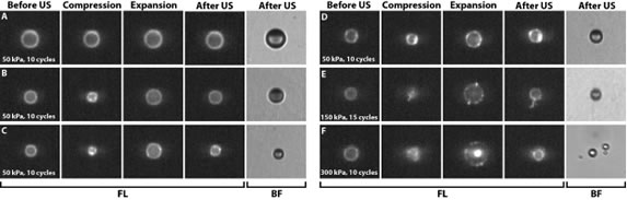
High speed fluorescence recordings with UPMC Cam reveal a variety of microbubble coating behaviors not evident from brightfield recordings. For microbubble with a homogeneous coating before ultrasound (50 kPa), we observed homogeneous fluorescence during oscillation (A), “hot spots” (focal areas of increased fluorescence intensity) only during compression (B), or hot spots during oscillation that persisted after US (C), suggesting that lipids can reach speeds on the order of 1 m/s. For microbubble with hot spots before US, the hot spots persisted or intensified (D). Occurrence of hot spots during ultrasound could be due to buckling. At higher acoustic pressures, we also observed other phenomena: formation of a tail (E) that was not evident in BF, or a non-continuous lipid coating during the expansion phase (F), suggesting a ruptured coating correlating with deflation. Adapted from Kooiman et al, IEEE IUS 2014.