Molecular Biology and Chemistry
The Center has state of the art laboratories to support a spectrum of molecular biologic and chemical techniques for developing our ultrasound-based applications. These include methods for protein characterization, plasmid synthesis, protein purification, protein synthesis (e.g. VEGF), and PCR. Our facilities permit the production of microbubbles using lipids and polymers, and incorporation of plasmid in the microbubble shell or targeting ligands on the microbubble surface. We can perform basic physical characterization of synthesized microbubbles, including determination of electrostatic charge, size distribution, and concentration. We have capabilities for sterile synthesis of microbubbles in a Clean Room specially designed for this purpose.
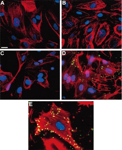
Above: Fluorescent micrographs of ECs after exposure to either nonspecific IgG–conjugated bubbles (A and B) or anti–ICAM-1–conjugated bubbles (C and D). Cytosolic F-actin is stained with rhodamine-phalloidin (red), nuclei are stained with Hoechst dye (blue), and microbubbles are labeled with fluorescein (green). Microbubbles have lost their gas and spherical shape during preparation for microscopy. A, Scale bar=10 µm. There was no adherence of nonspecific IgG–labeled bubbles to normal (A) or activated (B) cells. Although small numbers of anti–ICAM-1 bubbles adhered to normal ECs (C), numerous bubbles attached to activated ECs (D). Note intense rhodamine-phalloidin staining of F-actin indicative of activation (B and D) in cells exposed to IL-1ß. E, Enlargement of a single activated cell exposed to anti–ICAM-1 bubbles.
Source: Villanueva FS, Jankowski RJ, Klibanov S, Pina ML, Alber SM, Watkins SC, Brandenburger GH, Wagner WR. Microbubbles targeted to intercellular adhesion molecule-1 bind to activated coronary artery endothelial cells. Circulation. 1998 Jul 7; 98(1): 1-5.